THE RESEARCH SAYS
EP.
12
Can You Bond to Sclerotic Dentin?
Sclerotic dentin bonds weaker either way you etch it. Peumans' 2014 review and two 2026 air-abrasion studies show what actually helps: roughening, not brand.
THE RESEARCH SAYS
EP.
12
The verdict
ModerateBond strength to sclerotic dentin really is lower than to sound dentin. That part of the folklore holds up. What doesn't hold up is the idea that switching adhesive families fixes it: the largest reviews of non-carious cervical lesions find self-etch and etch-and-rinse producing statistically similar retention. What the evidence does support is mechanically opening the surface before bonding, extending your prep onto sound dentin at the margins, and (contrary to a lot of what we were taught) EDTA conditioning under a self-etch adhesive.
Moderate evidence: two systematic reviews, no dedicated RCT
The Class V that keeps coming back. It looks great at delivery, and eighteen months later the patient's back with a margin you can catch a fingernail on, or the whole restoration is in your suction trap. You blame flexure, or brushing, or a bruxer you should have splinted. Sometimes that's fair. A lot of the time the real problem was sitting in the prep the whole visit: sclerotic dentin, the glassy, hypermineralized substrate abfraction and erosion leave behind, and it does not bond like the dentin in your textbook photos.
Ask five dentists what to do about it and you'll get five confident, contradictory answers: roughen it, don't bother, use total-etch, use self-etch, condition with EDTA first, never use EDTA on this stuff. I went back to the primary literature to see which of those survive contact with an actual abstract. Two things surprised me. Some specific numbers I'd heard repeated for years (a roughening step said to more than double bond strength, a multi-year RCT on air abrasion) don't trace back to any indexed study I could find, and they aren't in this article. And a claim I'd have bet against, that EDTA helps rather than hurts sclerotic dentin, turned out to have the better paper trail.
The question
In adults with cervical lesions caused by dentin sclerosis (tubule occlusion and a hypermineralized surface, not a fresh caries excavation) does the bonding protocol (adhesive strategy, mechanical roughening, or chemical conditioning) change bond strength or clinical retention enough to matter? "Enough to matter" means a difference likely to change how many years the restoration survives, not a footnote in a lab report.
The mechanism
Healthy dentin is permeable: etch it, and acid opens the tubules and exposes collagen that resin infiltrates to form the hybrid layer and resin tags that do most of the retentive work. Sclerotic dentin is a defense response. Mineral occludes the tubules, and many lesions also carry a hypermineralized surface layer that resists both phosphoric acid and self-etch primers.4 That leaves two separate problems: often no open tubules for resin tags, and an acid-resistant layer sitting on top of what tubules remain.
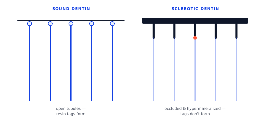 Illustrative diagram, not to scale. Mechanism described in 4.
What the evidence says
Peumans et al., 2014: systematic review of clinical trials
What they did. Reviewed randomized trials of adhesives in non-carious cervical lesions (1950–2013), 18+ months of follow-up, and calculated the average annual failure rate (AFR) per adhesive class.1
What they found. Glass ionomer and mild two-step self-etch had the lowest failure rates. Strong self-etch and ordinary two-step etch-and-rinse scored among the worst.1
Why it matters. This runs opposite to "etch-and-rinse always outperforms self-etch." Chemistry within a class mattered more than which broad family was chosen.
Schroeder et al., 2017: meta-analysis of adhesive strategy
What they did. Pooled 29 randomized trials comparing self-etch against etch-and-rinse in cervical lesions for retention, sensitivity, and discoloration.2
What they found. No significant difference in retention or postoperative sensitivity at any follow-up. Etch-and-rinse had less marginal discoloration at 18 months–2 years and 4–5 years.2
Why it matters. For keeping the restoration in the tooth, adhesive family looks close to irrelevant. For a margin that resists staining, etch-and-rinse has a modest edge.
Szesz et al., 2016: meta-analysis of selective etching
What they did. Reviewed trials of self-etch adhesives with and without selective enamel etching in cervical lesions, 1–5 years of follow-up.3
What they found. Selective etching improved marginal adaptation at nearly every interval and cut retention loss significantly at the 3-year mark.3
Why it matters. A narrower, more favorable read on selective etching than Peumans' pooled retention data: worth keeping in a self-etch protocol even where the broader retention signal is mixed.
Tay & Pashley, 2004: review of the substrate
What they did. The foundational review of sclerotic dentin's ultrastructure, synthesizing electron-microscopy and bond-strength studies.4
What they found. Beyond describing the occlusion and hypermineralized layer, the review notes that extending a restoration's margins onto peripheral sound dentin or enamel likely produces bond strength adequate to retain a Class V by adhesion alone.4
Why it matters. You may not fix the bond to sclerotic dentin itself much, but you can often change how much of the bonded surface is sclerotic versus sound tooth structure.
Karakaya et al., 2008: in vitro adhesive comparison
What they did. Bonded sclerotic and sound dentin with self-etch, total-etch, and glass-ionomer adhesives and measured microtensile bond strength.5
What they found. Self-etch outperformed the others on sound dentin; on sclerotic dentin, all three dropped and the difference between them disappeared.5
Why it matters. More evidence the substrate, not the adhesive brand, is doing most of the damage.
Chemical conditioning: Luque-Martínez, 2018, and Wang, 2019
What they did. Two in vitro studies tested EDTA conditioning before self-etch bonding on sclerotic dentin: one at 12 months,6 one also testing sodium hypochlorite combined with phosphoric acid.7
What they found. EDTA produced the highest immediate bond strength in both studies and, in Luque-Martínez's, resisted the degradation the non-EDTA groups showed at 12 months. Wang's phosphoric-acid-plus-hypochlorite groups matched or beat bond strength on untreated normal dentin.67
Why it matters. This directly reverses the teaching that EDTA and hypochlorite "wreck" the bond on sclerotic dentin. In these two studies, both did the opposite.
Mechanical roughening: two 2026 air-abrasion studies
What they did. Two recent in vitro studies tested aluminum-oxide air-particle abrasion before bonding to natural sclerotic dentin: one on premolars comparing sclerotic to non-sclerotic teeth from the same set,8 one on molars comparing three adhesives at 24 hours and 6 months.9
What they found. Air abrasion improved bond strength specifically on sclerotic dentin while reducing it on non-sclerotic dentin from the same teeth.8 Paired with an MDP-containing self-etch adhesive, it produced a 53% bond-strength increase over an untreated control, stable at 6 months.9
Why it matters. This is the closest thing in the current literature to a real number behind "roughening helps": substrate-specific, not a blanket rule, but quantified and durable at 6 months.
Evidence at a glance
| Study | Design | Sample | Follow-up | Finding | Weight |
|---|---|---|---|---|---|
| Peumans et al., 2014 | Systematic review | 42+ trials | 18 mo–5+ yr | Mild self-etch & GI lowest failure; etch-and-rinse among worst | High |
| Schroeder et al., 2017 | Meta-analysis | 29 trials | Up to 5 yr | No retention difference, self-etch vs. etch-and-rinse | High |
| Szesz et al., 2016 | Meta-analysis | 10 trials | 1–5 yr | Selective etching cut retention loss at 3 yr | Moderate |
| Tay & Pashley, 2004 | Narrative review | n/a | n/a | Sound-margin extension restores adequate bond | Moderate |
| Karakaya et al., 2008 | In vitro (µTBS) | 30 teeth | Immediate | Adhesive type stopped mattering on sclerotic dentin | Low |
| Luque-Martínez et al., 2018 | In vitro (µTBS) | 28 teeth | 12 mo | EDTA raised bond strength, slowed degradation | Low |
| Wang et al., 2019 | In vitro (µTBS) | 80 teeth | Immediate | Acid + hypochlorite, and EDTA, raised bond strength | Low |
| de Abreu et al., 2026 | In vitro (µTBS) | 48 teeth | Immediate | Air abrasion helped sclerotic, hurt sound dentin | Low |
| Del Cid Rodríguez et al., 2026 | In vitro (µTBS) | 16 teeth | 24 hr & 6 mo | Al₂O₃ + MDP self-etch: +53%, stable at 6 mo | Low |
What this means Monday morning
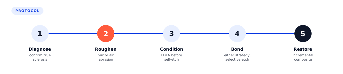 Illustrative protocol synthesized from the studies above, not a validated clinical guideline. Steps drawn from 12346789.
- Confirm it's actually sclerotic dentin: glassy, translucent, hardened margins with years of history, not a soft, active lesion.
- Roughen the surface before bonding: coarse diamond or aluminum-oxide air abrasion. Tied to higher bond strength on sclerotic dentin specifically, one study reporting a 53% gain.89
- Extend the prep onto sound peripheral dentin or enamel wherever the lesion allows it.4
- Pick an adhesive strategy and stop second-guessing it: self-etch and etch-and-rinse produce similar retention.12 Running self-etch, add selective enamel etching at the margins.3
- Consider EDTA conditioning under a self-etch adhesive on a lesion you expect to be difficult.67
- Restore with a well-adapted, incremental composite to manage shrinkage stress at a margin already working against you: my own habit, not a citation.
Where the evidence is thin
Almost everything here except the two clinical meta-analyses is in vitro: extracted teeth, no mouth, no years of function. The 2026 air-abrasion studies are small (48 and 16 teeth) and single-center, with no clinical follow-up yet. None of the clinical trials confirmed dentin sclerosis histologically case by case: "non-carious cervical lesion" and "sclerotic dentin" overlap but aren't identical. And a couple of numbers repeated for years in lectures (a roughening step said to more than double bond strength, a multi-year in-mouth RCT of air abrasion) don't trace to any real, indexed study I could find, so they're left out here. Nobody has run the trial that would settle this cleanly: isolate sclerotic dentin, randomize roughening versus none, follow patients for five years.
The bottom line
Sclerotic dentin bonds worse than sound dentin, full stop, and no adhesive family reliably closes that gap on its own. The evidence supports mechanically opening the surface, extending margins onto sound tooth structure, and (running self-etch) not being afraid of EDTA. Pick either major adhesive strategy with confidence and spend your effort on the surface instead.
References
- Peumans M, De Munck J, Mine A, Van Meerbeek B. Clinical effectiveness of contemporary adhesives for the restoration of non-carious cervical lesions. A systematic review. Dental Materials. 2014;30(10):1089-1103. DOIPubMed
- Schroeder M, Correa IC, Bauer J, Loguercio AD, Reis A. Influence of adhesive strategy on clinical parameters in cervical restorations: A systematic review and meta-analysis. Journal of Dentistry. 2017;62:36-53. DOIPubMed
- Szesz A, Parreiras S, Reis A, Loguercio A. Selective enamel etching in cervical lesions for self-etch adhesives: A systematic review and meta-analysis. Journal of Dentistry. 2016;53:1-11. DOIPubMed
- Tay FR, Pashley DH. Resin bonding to cervical sclerotic dentin: a review. Journal of Dentistry. 2004;32(3):173-196. DOIPubMed
- Karakaya S, Unlu N, Say EC, Ozer F, Soyman M, Tagami J. Bond strengths of three different dentin adhesive systems to sclerotic dentin. Dental Materials Journal. 2008;27(3):471-479. DOIPubMed
- Luque-Martinez IV, Muñoz MA, Hass V, Sutil E, Reis A, Loguercio AD. EDTA conditioning increases the long-term microtensile bond strength to sclerotic dentin mediated by self-etch adhesives. Journal of Adhesive Dentistry. 2018;20(5):397-403. DOIPubMed
- Wang J, Song W, Zhu L, Wei X. A comparative study of the microtensile bond strength and microstructural differences between sclerotic and normal dentine after surface pretreatment. BMC Oral Health. 2019;19(1):216. DOIPubMed
- de Abreu NMR, Ñaupari-Villasante R, Moreira PHA, Loguercio AD, de Sousa FB. Air-abrasion with aluminum oxide particles in non-carious cervical lesions: impact on sclerotic superficial layer, surface topography, and restorative adhesive performance. Journal of Esthetic and Restorative Dentistry. 2026 Aug 31. DOIPubMed
- Del Cid Rodríguez S, Carda Batalla C, Agustín-Panadero R, González-Angulo E, Román-Rodríguez JL. Influence of air-abrasion pretreatments and adhesive composition on bond durability to natural sclerotic dentin. Dentistry Journal. 2026;14(8):469. DOIPubMed
This article summarises published research for clinicians. It is not a substitute for your own reading of the studies or your clinical judgment.
Sclerosis is a defense response, and so is a lot of what shows up in a prep. See how that plays out elsewhere in Should You Leave Caries Behind? If patients ask why the margin feels different afterward, Does Gluma Reduce Post-Op Sensitivity In Dental Fillings? covers that question. And if a Class V keeps failing and you're tired of redoing it for free, Handling Dental Warranties is the business side of the same problem.
Questions
What dentists ask about this.
Can you get a reliable bond to sclerotic dentin?
Bond strength to sclerotic dentin really is lower than to sound dentin, and no adhesive family reliably closes that gap on its own. What helps is mechanically roughening the surface, extending the prep onto sound dentin, and (running self-etch) EDTA conditioning.
Does self-etch or etch-and-rinse adhesive bond better to sclerotic dentin?
Neither has a meaningful edge on retention. A meta-analysis of 29 randomized trials found no significant difference in retention or post-operative sensitivity between self-etch and etch-and-rinse in cervical lesions, though etch-and-rinse showed less marginal discoloration.
Does EDTA conditioning hurt or help bonding to sclerotic dentin?
It helps, reversing what many of us were taught. Two in vitro studies found EDTA produced the highest immediate bond strength on sclerotic dentin and resisted the degradation seen at 12 months in adhesive without it.
Does air abrasion improve bonding to sclerotic dentin?
Yes, specifically on sclerotic dentin: air abrasion improved bond strength there while reducing it on non-sclerotic dentin from the same teeth. Paired with an MDP-containing self-etch adhesive, one study found a 53% bond-strength increase over an untreated control, stable at 6 months.
Why is a Class V restoration on sclerotic dentin more likely to fail?
Sclerotic dentin is a hypermineralized defense response that occludes the dentinal tubules, leaving fewer open tubules for resin tags to form and an acid-resistant surface layer that resists both phosphoric acid and self-etch primers.
Key terms
From the reference library
- Scotchbond Universal (2011): the adhesive that created the universal category, and what the etching-mode evidence says3M · 2011
- Dentin hypersensitivity: the Canadian Advisory Board consensus (2003), a diagnosis of exclusion with a first-line treatment the trials never quite confirmedCanadian Advisory Board on Dentin Hypersensitivity · 2003
Where to next
Clinical judgement is half of it. The other half is the business around the chair.